Digitizing Collections
The Museum of Primatology (MOP) uses medical-grade scanners to produce 2D images of X-ray films and 3D volumetric density datasets of skeletal material. The resulting data is integrated into a web-accessible database, integrating the virtual skeletal collection with the power to perform advanced database searches to find data of interest online and the ability to view, download, and experiment with the digital data off-line. Creating a virtual skeletal collection preserves the physical specimens, avoiding wear and degradation caused by handling. And, once the materials have been digitized, annotated, and integrated into the web-accessible database, a number of exciting digital anthropogeny applications become possible.
2D X-Ray Film Scans
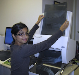
Medical X-ray film scanners are used to digitize high dynamic range (HDR) images from radiographic X-ray films capturing image resolutions of over 6 megapixels. This level of image quality in digital form eliminates the need to handle the original films. Many powerful software tools are available to enable digital anthropogeny techniques.
 |
 |
| X-Ray Film Digitizing | Digitized Radiograph Images |
Films are scanned and then annotated (including species, bone names, anatomical locations, and when available gender and age), resulting in fully annotated DICOM and JPEG image files. All of these digital radiographs may be searched by any annotated property within the web-accessible database (there are nearly 2000 radiograph images from our collections in the database).
3D CT Scans
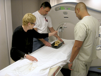
Clinical CT scanners are used to digitize larger specimens at 0.5mm resolution while Micro CT scanners are used to digitize smaller specimens at 9 micron resolution (fine enough to see and measure the trabecular wall thickness of bones). Many specialized software applications can be used for digital anthropogeny techniques.
 |
|
| CARTA's Pilot CT Scanning Session | CT Scan Slices |
Bones are scanned and then annotated (including species, bone names, anatomical locations, and when available gender and age), resulting in fully annotated DICOM and JPEG image files. All of these digital tomographs may be searched by any annotated property within the web-accessible database.
 |
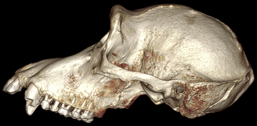 |
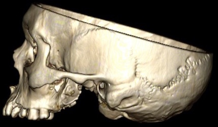
| Human Skull CT Rendering | Chimpanzee Skull CT Rendering |
The slices (see the slice video above) from the 3D tomographic data can be opened in medical imaging software and interactively rendered as one complete volumetric object (as the human and chimpanzee skulls above) and analyzed.
Digitizing Advantages
Preservation
- Physical specimens can be damaged over time with handling. Digital data can be provided to any number of users without any loss.
- Physical specimens inevitably deteriorate in storage (even without handling). Digital scans can be stored indefinitely without degradation.
- Digitizing preserves a snapshot (of the visual or density aspects) in time of the physical specimens.
- Digital scans can be sliced, diced, and otherwise processed and examined without destructive consequences to the physical specimen.
Access
- Digital scans of specimens can be electronically distributed so users do not have to travel to a physical location to use the collection for teaching or research.
- Images and data stored and annotated in a web-accessible database enable it to be searched to easily find what you need.
- Images and data can be downloaded to a user's computer for local off-line analysis and visualization.
Applications
- Enables the use of modern digital analysis and visualization techniques (see digital anthropogeny).
- Numerous teaching and training opportunities (see education).

