Digital Tools
The Museum's digital anthropogeny initiative, in conjunction with the San Diego Supercomputer Center, aims to modernize collection management, education, and research of anthropogenic materials through technological applications. The advantages enabled by integrating the collections database and digitizing technology allows the Museum of Primatology (MOP) to overcome limitations that would otherwise be problematic or impossible with the physical specimens alone. Students and researchers will benefit from digital anthropogeny tools such as powerful data search, data download, and off-line visualization and analysis functions.
Specimen Data Search
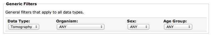
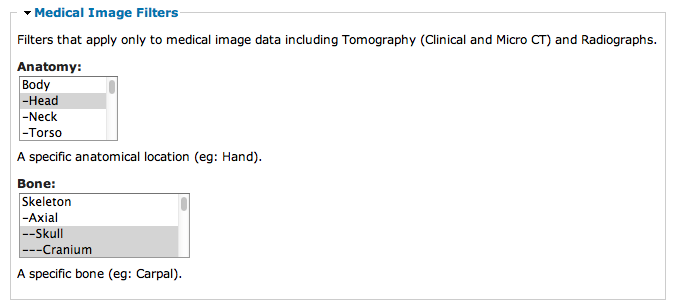
All of the physical specimens in the MOP catalog, as well as all of the digitized data, will be searchable by a rich set of attributes stored in the collections database. The scope of data search filters available to students and researchers spans general properties, such as species, sex, and age group, to type-specific criteria, including anatomical location, bone-specific conditions, and pathologies.
Specimen Data Access
The search results can be used in a variety of ways:
- Detailed attributes can be viewed for each specimen on detail pages by clicking on the search result links (includes online images of medical image data such as radiographs and CT volumes).
- The tabular search result metadata may also be exported (as CSV files) for offline analytics.
- For digitized data products, the raw data files (for example, ZIP archives containing DICOM medical image files for 2D radiographs and 3D CT volumes) can be downloaded for local visualization (using offline medical visualization software) and analysis (using spreadsheet or statistics packages).
Visualization and Analysis
Medical image data (such as 2D radiographs, Clinical CT, and Micro CT) can be downloaded and used for a number of applications in both education and research. Once the desired data is downloaded, MOP users can take advantage of many freely available visualization and analysis tools. Some medical exploration applications include:
- Examine external structures in order to compare form and function between species.
- Explore 2D cross-sections and measure internal structures in order to compare growth and development differences between species.
- Crop to reveal specific structural features (without the destructive consequences that the physical specimen would require).
- Interactively define transfer functions in order to hide less dense structures (like trabecular and cortical bone) while highlighting others (say, teeth).
- Easily export images and animations for presentations and publications.
Volumetric scans can be interactively viewed and explored in 3D using one of the free medical image applications (for example, Osirix).
Medical images data can be measured using length, area, and volume measuring tools.
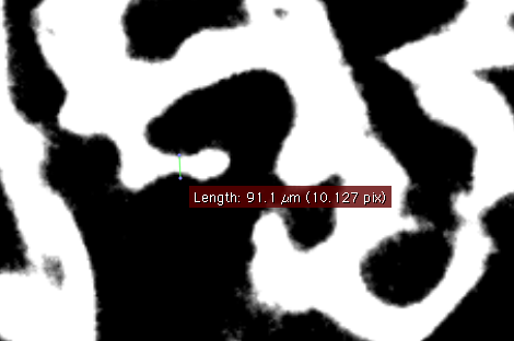
Medical imaging techniques are capable of making measurements more accurately than can be achieved using traditional measuring instruments. For example, MOP has used Micro CT scanning equipment to digitize bone specimens at 9 micron resolution (more than 2 orders of magnitude smaller than the best digital calipers can even measure!)
Using 3D visualization software, students and researchers may examine fine detail of internal structures using completely non-destructive interactive tools. Traditional techniques required permanent damage, such as the use of saws to remove a calotte for exploring internal brain cavity structures (as seen in the specimen above).

